41 dog muscle anatomy diagram
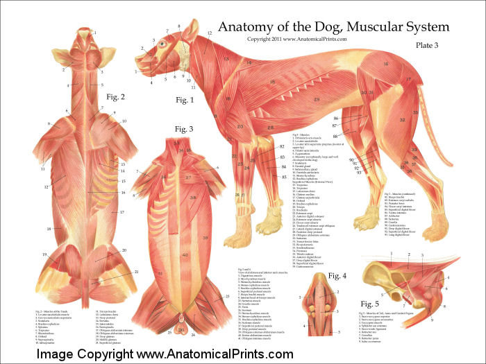
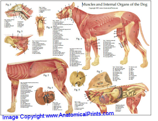
Dog Muscle Anatomy Diagram In this image, you will find scutularis, sternomastoideus, brachiocephalicus, trapezius, latissimus dorsi, iliocostalis, sartorius, tensor fasciae latae, glutaueus medius, crest of pelvis bone, glutaeus superficialis, trochanter major, semitendinosus, biceps femoris, quadriceps femoris, gastrocnemius, flexor hallucis ...
Dog front leg muscles anatomy You will find the extrinsic and intrinsic muscles in the front leg of a dog. I will show you the essential muscles from the dog front leg anatomy with a labeled diagram. Fine, if you want to know more about the dog muscle anatomy, you may read the article here.
Gain a comprehensive understanding of your dog's health with our veterinary guide to cat anatomy complete with diagrams, images and simple explanations. Introduction Anatomical terminology Pet senses Cardiovascular system - Heart & circulatory system Digestive system Musculoskeletal system Respiratory system Urogenital system - Urinary ...

Dog muscle anatomy diagram
A major part of a dog's anatomy is their musculature. This is a system formed by muscles, tendons and ligaments. A dog can have between 200 and over 400 muscles. Again, the amount of muscles an individual dog has depends on the breed and the individual. Curiously, some dog breeds will have more than 50 muscles in their ears alone.
Anatomy of the Canine Male Urogenital System (An Overview) Kidney. NAV Term: Ren . What is this? Each kidney is located dorsally in the abdominal cavity (in contact with the lumbar hypaxial muscles), and is retroperitoneal in position. The right kidney is slightly more cranial (at the level of lumbar vertebrae 1-3) than the left kidney (at the ...
The thoracic division of dog spine anatomy consists of the thoracic vertebrae, thick intervertebral discs, spinal cord, and thoracic spinal nerves. Now, I will show you some unique anatomical facts of the thoracic vertebrae of a dog so that you may identify it so quickly. You will find thirteen thoracic vertebrae in the vertebral dog column.
Dog muscle anatomy diagram.
Dog Muscle Anatomy Would you be surprised to know that a dog has 700 muscles in their body which make up 45% of their overall body weight. Muscles enable us to move. They stabilize our joints and maintain our posture; this is exactly the same for dogs.
The Muscle Anatomy of a Dog Pictured above is Ace from DarkDynastyK9s, arguably one of the most muscular dogs in the world. Ace's most predominant muscles are his triceps, Bicep Femoris, Scapular Deltoid, Acromion Deltoid, and his lower Trapezius. Ace uses Bully Max™ along with a 30/20 (30% Protein, 20% Fat) kibble.
For Practitioners. Practitioners and their clients benefit from EasyAnatomy's interactive canine model and animations of common pathologies. As the most advanced interactive 3D canine anatomy client communication tool, EasyAnatomy breaks down the communication barrier and increases client compliance. Learn more.
Dog anatomy poster created using vintage images. The poster shows the superficial muscles, skeletal system with surface anatomy. Superior view of the spinal column with musculature for veterinary acupuncture and chiropractic.
The external anatomy of a dog is quite simple to understand. The following diagram and paragraph attempt to explain it in brief. The muzzle is of varying lengths, depending on the breed. Whiskers, present on the muzzle, are of some sensory use. Dogs also have a 'stop' on their heads, which is the point where the muzzle ends and the forehead begins.
Anatomy of Dog (thoracic muscles) Superficial pectoral Deep pectoral sternothyroideus Omotransversarius 2 parts: descending and transverse... O: first three sternebrae/… O: ventral part of sternum/median fiborus raphe between adjace… covered by sternohyoideus at origin... O: first costal cartilage… deeper than cleidocephalicus...
Understanding and knowing your dog's leg anatomy will help learn the possible weaknesses, injuries, and the best ways how to treat them. The dog is carried around by the forelegs and the hind legs. Much as the hind legs have got larger muscles which make them stronger, they only carry around one-third of its body weight.
Anatomy is at the core of the veterinary profession. ... and inserts at the deltoid tuberosity of the humerus at number 3 in the diagram. The supraspinatus muscle does the opposite function to the deltoid, it is the main shoulder extensor (increases the angle between two body parts) and inserts on the greater tuberosity at number 4. The humerus ...
The pictures in this section are reprinted with permission by the copyright owner, Hill's Pet Nutrition, from the Atlas of Veterinary Clinical Anatomy. These illustrations should not be downloaded, printed or copied except for personal, non-commercial use.
Dog anatomy details the various structures of canines eg. Neck Cancer Anatomy Headandneckcancerguide Org All dogs and all living canidae have a ligament connecting the spinous process of their first thoracic or chest vertebra to the back of the axis bone second cervical or neck bone which supports the weight of the head without active muscle ...
Dog Joint Anatomy The anatomy of dogs varies tremendously from breed to breed, more than in any other animal species, wild or domesticated. Yet there are physical characteristics that are identical among all dogs, from the chihuahua to the giant Irish wolfhound. They have small, tight feet, walking on their toes; their
Anatomy of the dog - Illustrated atlas This modules of vet-Anatomy provides a basic foundation in animal anatomy for students of veterinary medicine. This veterinary anatomical atlas includes selected labeling structures to help student to understand and discover animal anatomy (skeleton, bones, muscles, joints, viscera, respiratory system ...
Dogs have a third trochanter, which is the attachment site of the superficial gluteal muscle. Canine medial and lateral femoral condyles are equally prominent, but the articular surface of the medial femoral condyle projects more cranially than that of the lateral femoral condyle. There are three sesamoid bones in the caudal stifle joint region.
Dog anatomy comprises the anatomical studies of the visible parts of the body of a domestic dog.Details of structures vary tremendously from breed to breed, more than in any other animal species, wild or domesticated, as dogs are highly variable in height and weight. The smallest known adult dog was a Yorkshire Terrier that stood only 6.3 cm (2.5 in) at the shoulder, 9.5 cm (3.7 in) in length ...
Labeled anatomy of the head and skull of the dog on CT imaging (bones of cranium, brain, face, paranasal sinus, muscles of head) This module of vet-Anatomy presents an atlas of the anatomy of the head of the dog on a CT. Images are available in 3 different planes (transverse, sagittal and dorsal), with two kind of contrast (bone and soft tissues).
2] cardiac muscle = striated; musculature of the heart 3] skeletal muscle = striated; generally attached to bone; usually under voluntary control Skeletal Muscle Skeletal (striated) muscle is composed of elongate, multinucleated cells (muscle fibers). Different types of muscle fibers are found among the various skeletal muscles of the body, e.g.,
14,382 dog anatomy stock photos, vectors, and illustrations are available royalty-free. See dog anatomy stock video clips. of 144. stomach dog muscle and bones animal animal organs dog liver dog internal organs dog muscle anatomy dog lungs dog spine canine anatomy dog digest. Try these curated collections.
The aforementioned layers are functional layers of dog eye anatomy. The functionality of the eye is made possible with various anatomical features, which includes the, eyelids, eyelashes, conjunctiva, cornea, iris, pupil, lens, third eyelid (nictitans gland) lacrimal gland and viteous chamber of the eye.
Canine Muscle Origins, Insertions, Actions and Nerve Innervations The purpose of this document is to provide students of canine anatomy a simple reference for muscular origins, insertions, actions and nerve innervations without having to search through the overwhelming verbiage that accompanies most canine anatomy texts. Millerʼs Anatomy of
Anatomy of Dog (thoracic muscles) Superficial pectoral Deep pectoral sternothyroideus Omotransversarius 2 parts: descending and transverse... O: first three sternebrae/… O: ventral part of sternum/median fiborus raphe between adjace… covered by sternohyoideus at origin... O: first costal cartilage… deeper than cleidocephalicus...
Anatomy 1 Canine Muscles Diagram Quizlet. Dog Anatomy Muscular System Stock Image Z932 0463 Science Photo Library. Dog Veterinary Muscle Anatomy Poster 24 X 36 Zazzle. Muscle Anatomy Of The Dog Quiz. Dog Muscle Anatomy Diagram. Anatomy Of The Dog Bones Muscles 1 Chart Bullitor Acupuncture Charts For Horses And Dogs.
Dog Muscle Skeletal Anatomy Poster - 18" X 24" Custom designed veterinary poster. Printed on heavy weight HP satin finish paper. Available laminated. Dog anatomy poster. The poster shows the superficial muscles, skeletal system with surface anatomy. Superior view of the spinal column with musculature. Shipping UPS to USA addresses.
Dog muscular system diagram In this image, you will find dog, dog muscle anatomy, dog muscular system, cleidobrachialis, trapezius, latissimus dorsi, gluteal muscles, tensor fascia lata, biceps femoris, fascia lata covering vastus muscles in it.


0 Response to "41 dog muscle anatomy diagram"
Post a Comment