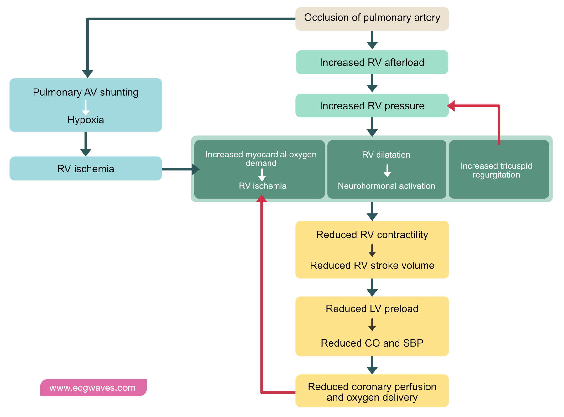
37 pulmonary embolism pathophysiology diagram
Acute pulmonary embolism 1: pathophysiology, clinical ... Greenspan RH. Pulmonary angiography and the diagnosis of pulmonary embolism. Prog Cardiovasc Dis. 1994 Sep-Oct; 37 (2):93-105. [Google Scholar] Hull RD, Raskob GE, Ginsberg JS, Panju AA, Brill-Edwards P, Coates G, Pineo GF. A noninvasive strategy for the treatment of patients with suspected pulmonary embolism. Pathophysiology of pulmonary embolism | McMaster ... Venous thromboembolism (VTE) » Pathophysiology of pulmonary embolism. Venous thromboembolism (VTE) » Pathophysiology of pulmonary embolism. Posted September 26, 2012 by Eric Wong.
Pulmonary Embolism (PE) - Lung and Airway Disorders ... Pulmonary Embolism. Pulmonary embolism is the blocking of an artery of the lung (pulmonary artery) by a collection of solid material brought through the bloodstream (embolus)—usually a blood clot (thrombus) or rarely other material. Pulmonary embolism is usually caused by a blood clot, although other substances can also form emboli and block ...
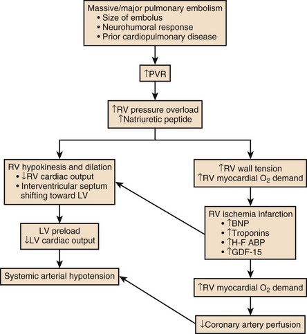
Pulmonary embolism pathophysiology diagram
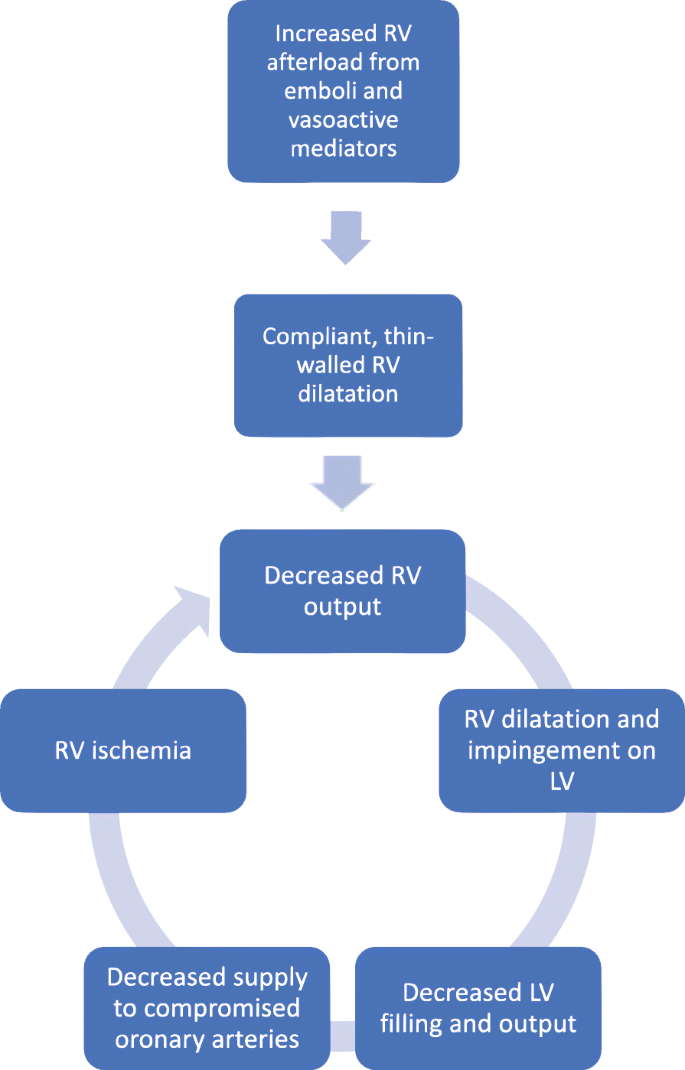
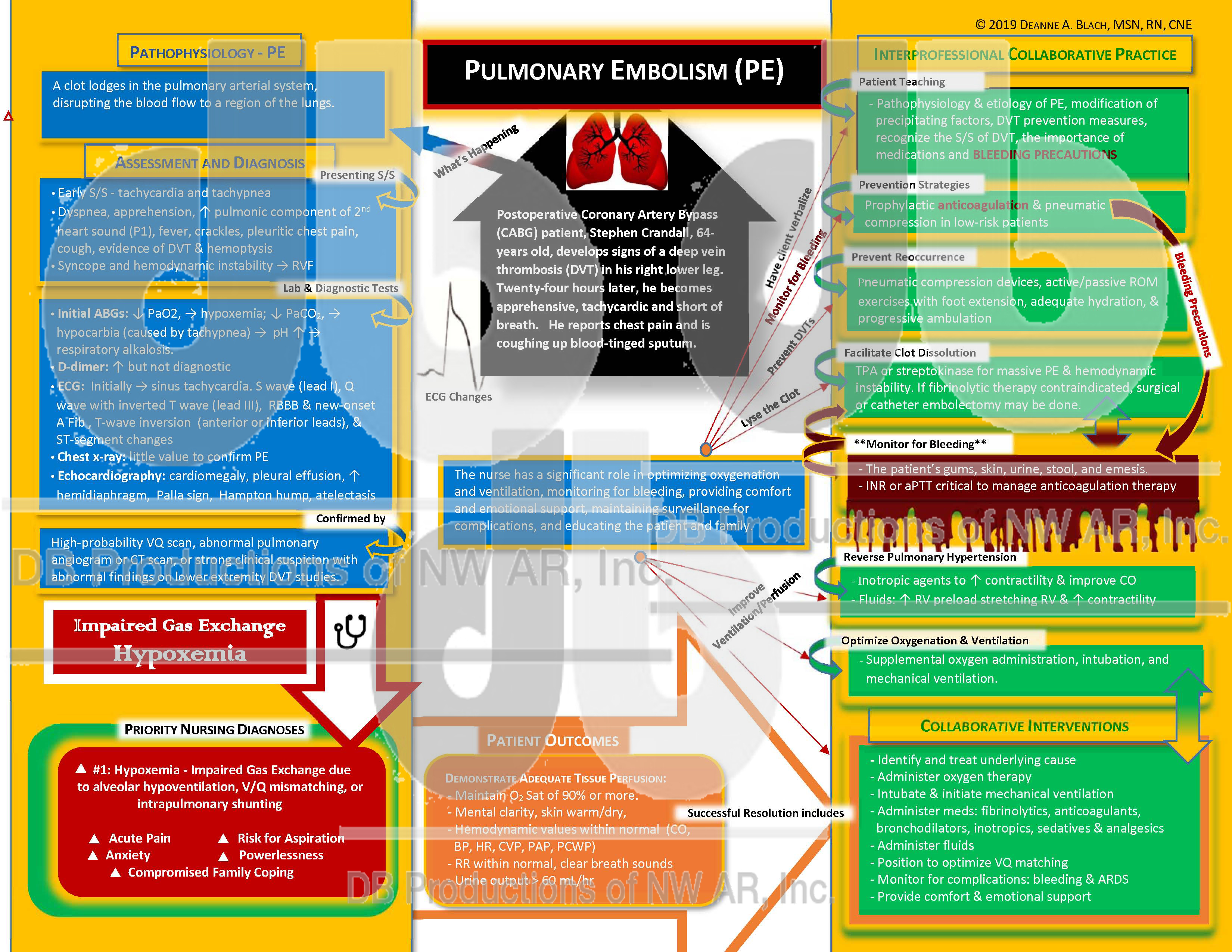
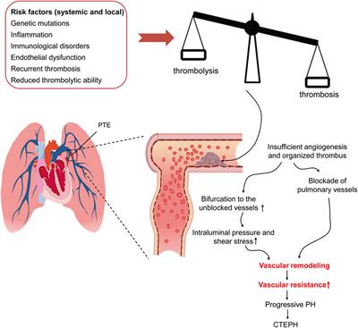
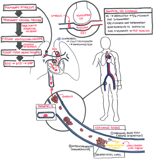
Pathophysiology | Pulmonary Embolism - Ohio State University PATHOPHYSIOLOGY. If there is an occlusion or partial occlusion of the pulmonary artery or its branches, it will cause a pulmonary embolism. Common cause: An embolized clot from deep vein thrombosis (DVT) involving the lower leg. Less common causes: Tissue fragments; Lipids; Foreign body; Air bubble; Amniotic fluid; Risk Factors Pathophysiology | Background information | Pulmonary ... What is the pathophysiology? Pulmonary embolism (PE) occurs when one or more emboli, usually arising from a thrombus (blood clot) formed in the veins, are lodged in and obstruct the pulmonary arteries. When a PE is present, the lung tissue is ventilated but not perfused, resulting in an intra-pulmonary dead space and impaired gas exchange ... Acute pulmonary embolism 1: pathophysiology, clinical ... Thrombotic pulmonary embolism is not an isolated disease of the chest but a complication of venous thrombosis. Deep venous thrombosis (DVT) and pulmonary embolism are therefore parts of the same process, venous thromboembolism. Evidence of leg DVT is found in about 70% of patients who have sustained a pulmonary embolism; in most of the remainder, it is assumed that the whole thrombus has ...
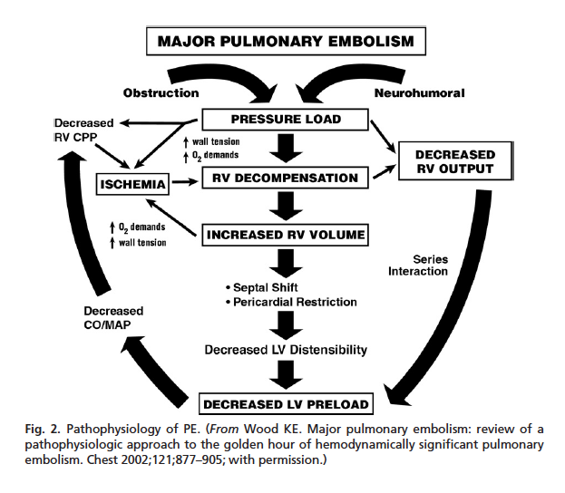
Pulmonary embolism pathophysiology diagram. PDF Acute pulmonary embolism 1: pathophysiology, clinical ... Acute pulmonary embolism 1: pathophysiology, clinical presentation, and diagnosis Martin Riedel German Heart Center, Munich, Germany Table 1 Risk factors for venous thromboembolic disease Venous stasis or injury,secondary hypercoagulable states: Immobilisation or other cause of venous stasis—for PDF Pulmonary Embolism - American Thoracic Society A pulmonary embolism (PE) is a blood clot that gets into blood vessels in the lungs and prevents normal flow of blood in that area. This blockage causes problems with gas exchange. Depending on how big a clot and number of vessels involved, it can be a life-threatening event. Pulmonary Embolism Left atrium Left ventricle Epidemiology, Pathophysiology, Stratification, and Natural ... Pulmonary embolism (PE) is a common and potentially fatal form of venous thromboembolism that can be challenging to diagnose and manage. PE occurs when there is obstruction of the pulmonary vasculature and is a common cause of morbidity and mortality in the United States. A combination of acquired a … Pulmonary edema: pathophysiology and diagnosis Two main types of pulmonary edema are recognized: first, cardiogenic (or hydrostatic) pulmonary edema from, as the name implies, an elevated pulmonary capillary pressure from left-sided heart failure; second, noncardiogenic (increased permeability) pulmonary edema from injury to the endothelial and (usually) epithelial barriers.
Pulmonary Embolism Nursing Care and Management: Study Guide Pathophysiology of Pulmonary Embolism. Click the image to enlarge. Obstruction. When a thrombus completely or partially obstructs the pulmonary artery or its branches, the alveolar dead space is increased. Impairment. The area receives little to no blood flow and gas exchange is impaired. Pulmonary embolism An update - PubMed Background: Pulmonary embolism is a common condition and can be the source of significant morbidity and mortality. Objective: This article reviews the approach to the diagnostic assessment and management of patients with suspected pulmonary embolism. Discussion: Various clinical decision rules and algorithms are available to assist in the diagnosis of pulmonary embolism, and the Wells score ... Pulmonary Embolism (PE) - Pulmonary Disorders - Merck ... Pulmonary embolism (PE) is the occlusion of pulmonary arteries by thrombi that originate elsewhere, typically in the large veins of the legs or pelvis. Risk factors for pulmonary embolism are conditions that impair venous return, conditions that cause endothelial injury or dysfunction, and underlying hypercoagulable states. Pulmonary embolism - PubMed Central (PMC) Pulmonary embolism (PE) is responsible for approximately 100,000 to 200,000 deaths in the United States each year. With a diverse range of clinical presentations from asymptomatic to death, diagnosing PE can be challenging. Various resources are available, ...
Pulmonary Embolism - SlideShare Pulmonary Embolism • Occlusion of a pulmonary artery (ies) by a blood clot. • Results from DVTs that have broken off and travelled to the pulmonary arterial circulation. • PE is one of the leading causes of preventable deaths in hospitalized patients. 20/01/20164. 5. 20/01/20165. 6. Pulmonary embolism pathophysiology - wikidoc Pulmonary embolism (PE) occurs when there is an acute obstruction of the pulmonary artery or one of its branches. It is commonly caused by a venous thrombus that has dislodged from its site of formation and embolized to the arterial blood supply of one of the lungs. The process of clot formation and embolization is termed thromboembolism. Acute Pulmonary Embolism - StatPearls - NCBI Bookshelf Pulmonary embolism (PE) occurs when there is a disruption to the flow of blood in the pulmonary artery or its branches by a thrombus that originated somewhere else. In deep vein thrombosis (DVT), a thrombus develops within the deep veins, most commonly in the lower extremities. PE usually occurs when a part of this thrombus breaks off and enters the pulmonary circulation. Pulmonary vascular disease Notes: Diagrams & Illustrations ... Chapter 129 Pulmonary Vascular Disease Figure 129.5 The histological appearance of pulmonary edema. PULMONARY EMBOLISM osms.it/pulmonary-embolism PATHOLOGY & CAUSES Blockage of pulmonary artery by a substance brought there via bloodstream Thrombus in remote site embolizes → lodges in pulmonary vascular tree → "pulmonary embolism" Obstruction of blood flow distal to embolism ...
Pathophysiology and causes of pulmonary embolism - Oxford ... Pulmonary embolus is predominantly due to thrombus breaking off from deep veins or from within the right heart, lodging within large or small vessels within the pulmonary vasculature, causing a variable degree of clinical features ranging from asymptomatic through to shock and cardiac arrest. Non-thrombotic causes include air or fat embolism. Outcome is predicated by the degree of right ...
Cor Pulmonale - StatPearls - NCBI Bookshelf The pathophysiology of cor pulmonale is a result of increased right-sided filling pressures from pulmonary hypertension that is associated with diseases of the lung. [5] [6] [7] Under normal physiologic conditions, the right ventricle pumps against a low-resistance circuit.
Acute pulmonary embolism 1: pathophysiology, clinical ... Thrombotic pulmonary embolism is not an isolated disease of the chest but a complication of venous thrombosis. Deep venous thrombosis (DVT) and pulmonary embolism are therefore parts of the same process, venous thromboembolism. Evidence of leg DVT is found in about 70% of patients who have sustained a pulmonary embolism; in most of the remainder, it is assumed that the whole thrombus has ...
Pathophysiology | Background information | Pulmonary ... What is the pathophysiology? Pulmonary embolism (PE) occurs when one or more emboli, usually arising from a thrombus (blood clot) formed in the veins, are lodged in and obstruct the pulmonary arteries. When a PE is present, the lung tissue is ventilated but not perfused, resulting in an intra-pulmonary dead space and impaired gas exchange ...
Pathophysiology | Pulmonary Embolism - Ohio State University PATHOPHYSIOLOGY. If there is an occlusion or partial occlusion of the pulmonary artery or its branches, it will cause a pulmonary embolism. Common cause: An embolized clot from deep vein thrombosis (DVT) involving the lower leg. Less common causes: Tissue fragments; Lipids; Foreign body; Air bubble; Amniotic fluid; Risk Factors














0 Response to "37 pulmonary embolism pathophysiology diagram"
Post a Comment