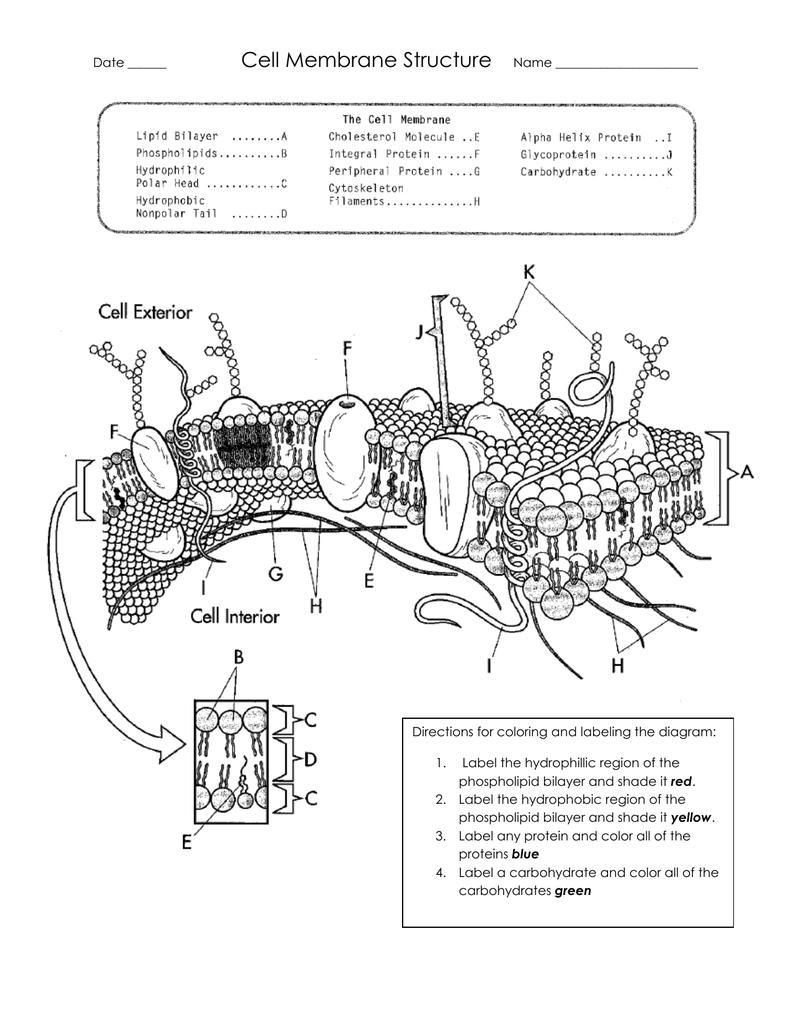
37 labeled diagram of plasma membrane
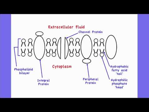
The plasma membrane of a corpuscle is a arrangement of lipids and proteins that forms the abuttals amid a cell's capacity and the alfresco of the cell. Plasma Membrane Structure. Corpuscle membrane abundant diagram. Cell Membrane or Plasma Membrane. The corpuscle membrane is a attenuate band fabricated up of proteins, lipids, and fats. All cells in animal body tissues are electrically polarized – in other words, they maintain a voltage difference across the cell's plasma membrane, known as the membrane potential. This electrical polarization results from a complex interplay between protein structures embedded in the membrane called ion pumps and ion channels .
18.04.2020 · As plasma is filtered from the glomerulus into Bowman’s capsule it travels through 3 layers, glomerular capillary endothelial layer, glomerular basement membrane, and Bowman’s capsule podocytes. The endothelial layer of the capillaries have small pores, called fenestrae, that allow water and soluble substances to exit the capillary.

Labeled diagram of plasma membrane
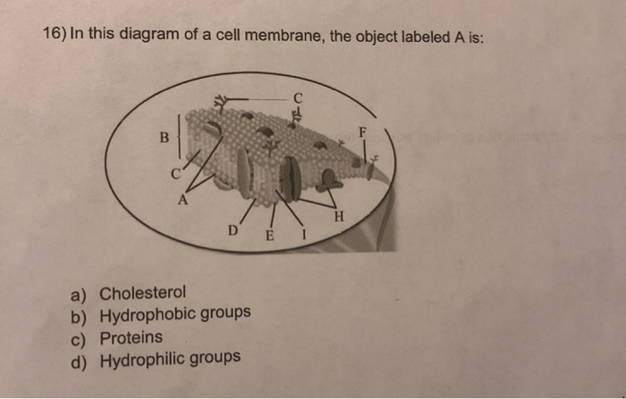
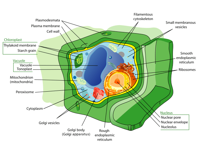
Presence of plasma membrane 2. The structure labeled A is present in all prokaryotic and eukaryotic cells. The structure is Ribosomes Cell wall Cell membrane Endoplasmic reticulum 3. The organelle labeled F is responsible for life in this planet and animal cells lack this organelle. The organelle is Mitochondrion Ribosomes Chloroplast Nucleus 4. This is the physical basis of … Diagram Of Animal Cell. Animal cells are eukaryotic cells that contain a membrane-bound nucleus. They are different from plant cells in that they do contain cell walls and chloroplast. The animal cell diagram is widely asked in Class 10 and 12 examinations and is beneficial to understand the structure and functions of an animal. 21.10.2021 · 4) Cell Membrane or Plasma Membrane. It is a thin, biological membrane having a thickness of 7.5-10 nm that separates the interior of the cell from the outside environment. The plasma membrane is selectively permeable in nature, which is mainly composed of lipids and proteins, with some carbohydrates attached to them. Functions
Labeled diagram of plasma membrane. 01.12.2021 · Figure: Diagram of the cell (plasma) membrane. Source: Wikipedia Structure of the plant cell (plasma) membrane. This is a bilipid membrane that is made up of protein subunits and carbohydrates, with a characteristic semi permeability factor. It surrounds the cell cytoplasm, thus enclosing its content. Functions of the plant cell (plasma) membrane Label Plasma Membrane. Here are a number of highest rated Label Plasma Membrane pictures upon internet. We identified it from well-behaved source. Its submitted by government in the best field. We tolerate this nice of Label Plasma Membrane graphic could possibly be the most trending topic afterward we share it in google benefit or facebook. Volvox Diagram Also see: MCQs on Volvox MCQs on Algae Volvox Characteristics A single colony of volvox looks like a ball of ~0.5 mm in diameter The plant body of volvox is a hollow sphere called coenobium, thousands of cells are arranged in the periphery of the sphere The cells of coenobium are of two types, germ cells and flagellated somatic cells 1-1 A generalized flow diagram of data ... The Plasma Membrane: Structure and Functions Figure 3.1 is a diagram of a portion of a plasma membrane. Select four differ- ent colors and color the coding circles and the corresponding structures in the diagram. Then respond to the questions that follow, referring to Figure 3.1 and inserting your answers in the answer blanks. Phospholipid ...
Bacteria diagram also indicates Periplasmic space, that is a cellular compartment discovered purely in bacteria that have an outer membrane and a plasma membrane . Game Statistics - Label the structure of the cell-surface … (Lucas Washington) This structure has two layers, and is represented in the diagram below. Membrane loss - both outer acrosomal membrane and plasma membrane are lost by forming vesicles during acrosome reaction. Acrosome reaction has a slow and rapid component: [30] Rapid - (seconds) efflux of calcium from intracellular stores, triggers fusion pores opening and the release of hybrid vesicles. However, the cell membrane in plant cells is quite rigid, while, the cell membrane in animal cells is quite flexible. As observed in the labeled animal cell diagram, the cell membrane forms the confining factor of the cell, that is it envelopes the cell constituents together and gives the cell its shape, form, and existence. Membranes 2.4.1 Draw and label a diagram to show the structure of membranes.. Figure 2.4.1 - Annotated drawing of a cell membrane. 2.4.2 Explain how the hydrophobic and hydrophilic properties of phospholipids help to maintain the structure of cell membranes.. Phospholipid molecules make up the cell membrane and are hydrophilic (attracted to water) as well as hydrophobic (not attracted to water ...
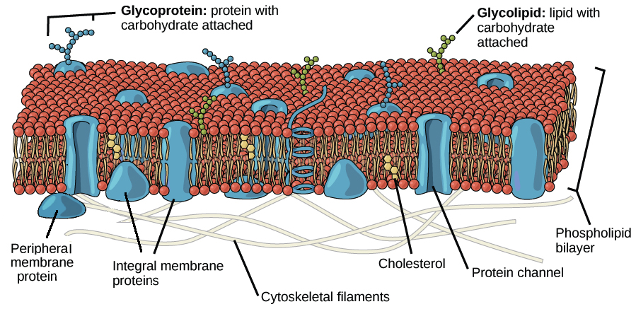
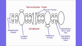
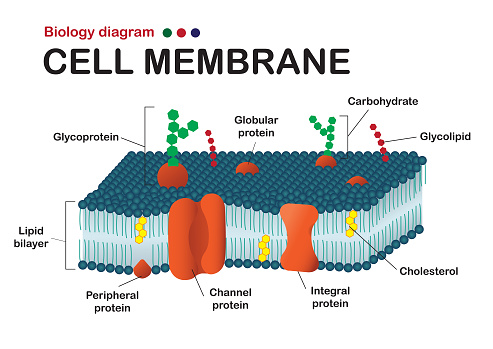
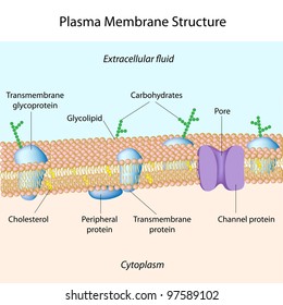
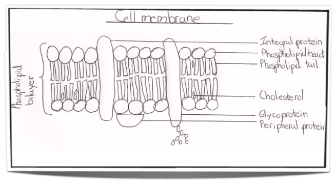
Early endosomes (E - labeled for EGFR, 5 minutes after internalisation, and transferrin), late endosomes/MVBs (M) and lysosomes (L) are visible. Bar, 500 nm. Endosomes are a collection of intracellular sorting organelles in eukaryotic cells. They are part of endocytic membrane transport pathway originating from the trans Golgi network. Molecules or ligands internalized from the … How To Draw And Label Golgi Apparatus Step By Step Tutorial Youtube Biology Diagrams Science Diagrams Body Diagram . Schematic Diagram Of A Cell Membrane Plasma Membrane Cell Membrane Cell Membrane Structure . Diffusion Osmosis And Active Transport Worksheet Science Notebook Rubric Osmosis Biology Worksheet . Define facilitated diffusion. movement of particles from an area of high to low concentration across a partially permeable membrane via protein channels or carriers. list five factors that affect the rate of simple diffusion. Temperature, diffusion distance, surface area, size of diffusing molecule, concentration gradient. Plasma Membrane - Components. It is composed of the following constituents: Phospholipids - forms the ultimate fabric of the membrane. Peripheral proteins - present on the outer or inner surface of phospholipid bilayer but are not implanted in the hydrophobic core. Cholesterol - folded between the hydrophobic tails of phospholipid membrane.
Structure of Plasma membrane | hasanul007's Blog
Jul 01, 2019 · The long-term circulatory effect of erythrocytes is mediated by a series of membrane proteins on the membranes surface. Among these, CD47 constitutes an integral membrane protein with five membrane-spanning regions, firmly embedding it in RBCMs, along with an IgV-like extracellular domain that contributes to RBCM survival in circulation 30.

Structure of the plasma membrane (article) | Khan Academy
Cell Membrane ColoringSKETCH AND LABEL a phospholipid coloring the heads red and the tails blue. Membrane worksheet name 1. Labeled Diagram Of Plasma Membrane Luxury In The Cell Membrane Plasma Membrane Phospholipid Bila Plasma Membrane Cell Membrane Cell Membrane Coloring Worksheet Help students visualize the structure of a cell membrane by using an analogy.
Structure of the plasma membrane (article) | Khan Academy
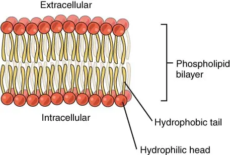
Identify the labeled parts of the plasma membrane below from the diagram. C A phospholipid phospholipid bilayer hydrophilic head hydrophobic tail D. check_circle.

Label Cell Membrane Diagram | Quizlet
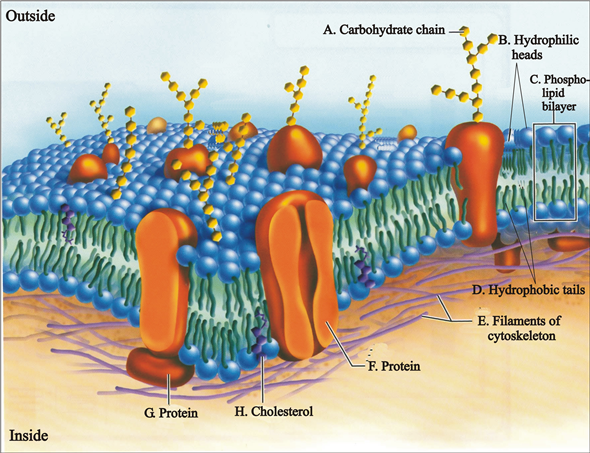
The principal components of the plasma membrane are lipids (phospholipids and cholesterol), proteins, and carbohydrate groups that are attached to some of the lipids and proteins. A phospholipid is a lipid made of glycerol, two fatty acid tails, and a phosphate-linked head group. Biological membranes usually involve two layers of phospholipids ...

Describe the fluid mosaic model of plasma membrane.
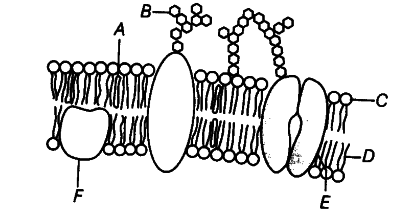
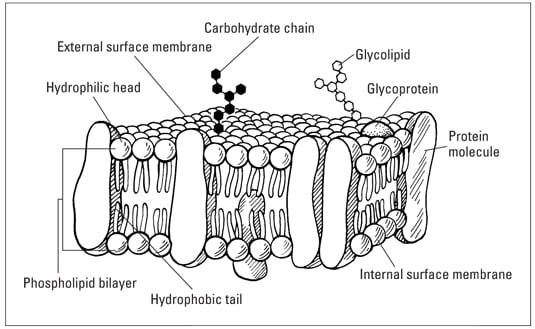
Label this diagram of the plasma membrane with the following terms: carbohydrate chain cholesterol cytoskeleton filaments glycolipid glycoprotein phospholipid bilayer protein molecule The sandwich model and its associated unit membrane model preceded the currently accepted fluid-mosaic model of membrane structure.
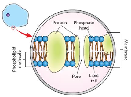
Diagram The Plasma Membrane And Label Each Component - Wiring ...
LAB #4- Plasma Membrane. STUDY. Learn. Flashcards. Write. Spell. Test. PLAY. Match. Gravity. Created by. Neville_Crick TEACHER. Terms in this set (11) Cholesterol. a lipid which stiffens phospholipid layers to provide support making it less fluid, less permeable and lowers freezing point of phospho lipid bilayer so cells do not freeze.

1 - nandin123
extocytosis. fusion of vesicles with the plasma membrane. vesicle membrane and membrane fuse. its used to export carbohydrates and proteins. endocytosis. cell takes in molecules to form vesicles from plasma membrane. phagocytosis. cell engulfs particle to be digested in food vacuoles.

Membranes
Plasma Membrane: 1. Living boundary of every cell. This is called unit membrane, plasmalemma, or cell membrane. 2. Made up of lipids and protein mainly. 3. The most accepted structure is called the ‘fluid mosaic’ model, proposed by Singer and Nicolson. 4. Being selectively permeable regulates the movement of molecules. Nucleus: 1. The brain ...
Cell/Plasma Membrane Structure Color & Label Perfect for Interactive Notebooks!
The components of the membrane are not stationary as is a plasma membrane diagram. Rather, they float freely through the membrane. ... 11. protein type that spans the plasma membrane Activity 2 ...

1.3 Mr. McGee, IB Biology (HL) - ppt video online download
Many different microscopic organisms can be found in pond ecosystems, including the three organisms shown in the diagram below. The primary cellular structures in each of these single-celled organisms are labeled in the diagrams. Some of the structures are common to all three organisms and the other structures are not.
2.4.1 Draw and label a diagram to show the structure of ...
In this article we will discuss about the structure of plasma membrane. This will also help you to draw the structure and diagram of plasma membrane. 1. The membrane which bounds the protoplasm (Fig. 292) of the cell of all living organisms including plants and animals is known as plasma membrane or cell membrane or plasmalemma.
Solved 16) In this diagram of a cell membrane, the object ...
The Phospholipid Bilayer. The plasma membrane is the most thoroughly studied of all cell membranes, and it is largely through investigations of the plasma membrane that our current concepts of membrane structure have evolved. The plasma membranes of mammalian red blood cells (erythrocytes) have been particularly useful as a model for studies of membrane structure.
Cell membrane christmas - Teaching resources
28.11.2021 · The plasma membrane is continuous with the plasma membrane of the cell and has the same phospholipid bilayer. The cytoplasmic sleeve is continuous with the cytosol that allows the exchange of materials between two cells. Desmotubule which is a part of the endoplasmic reticulum that provides a network between two cells and allows the transport of …
Biology Diagram Show Structure Of Cell Membrane Stock ...
Aug 02, 2021 · Cell Membrane. Every cell has a cell membrane, whether it be a plant or animal. A cell membrane is a division between the outside environment and the inside protoplasm of the cell.
Study Guide The Plasma Membrane
Essentially, the plasma membrane refers to the cell membrane that defines the boundaries of a Animal Cell Diagram Labeled Workshe Parts and Organelles of an Animal Cell in Cross Section Diagram Worksheet Colored Version.
Plant Cells: Labelled Diagram, Definitions, and Structure
Label this diagram of the plasma membrane. Step-by-step solution. Step 1 of 5. Plasma membrane contains the following parts: a) Glycoprotein: Glycoproteins are formed by covalent binding of oligosaccharide chains with a proteins present in plasma membrane. Chapter 5, Problem 11TY is solved.
Plasma membrane Images, Stock Photos & Vectors | Shutterstock
draw a labelled diagram of the fluid mosaic model of the plasma membrane - Biology - TopperLearning.com | 98ytnpi99. Practice Test - MCQs test series for Term 2 Exams. ENROLL NOW.
:max_bytes(150000):strip_icc()/cell-membrane-373364_final-5b5f300546e0fb008271ce52.png)
Cell Membrane Function and Structure
S1A shows that the two labeled fluorophores on H1 or H2 for signal reporting are heat resistant with relatively stable luminescence after ... The NTA particle size distribution diagram (Fig. 5A) shows that 91.5% of the sample EVs are distributed within 30–200 nm with the mean size of 137.8 nm, which accords with the exosomes subtype size range. The total EVs concentration …
Solved: Use these terms to label the following diagram of the ...
Some of the major parts of the plasma membrane are : phospholipids, glycolipids, cholesterol, integral membrane proteins (including transmembrane proteins), and peripheral membrane proteins. Jenny Norris. ... Fluid Mosaic Model Vector Illustration Labeled Stock Vector (Royalty Free) 1509614921.
IB Biology Notes - 2.4 Membranes
Membrane Proteins Can Be Associated with the Lipid Bilayer in Various Ways. Different membrane proteins are associated with the membranes in different ways, as illustrated in Figure 10-17.Many extend through the lipid bilayer, with part of their mass on either side (examples 1, 2, and 3 in Figure 10-17).Like their lipid neighbors, these transmembrane proteins are …
Cell Membrane Explained: Here's Everything You Need to Know ...
Aug 24, 2021 · The result showed that after a treatment of the cells with PS, which increases the content of acidic lipids in the plasma membrane 27, a higher FRET efficiency was detected compared to the solvent ...
Structure and Function of the Plasma Membrane by A-Thom-ic ...
Plasma, the liquid component of blood, comprises 55 percent of the total blood volume. It can separated by artificially spinning or centrifuging the blood at high rotations of 3000 rpm or higher. The blood cells and platelets that make up about 45 percent of the blood are separated by centrifugal forces to the bottom of a specimen tube, leaving ...

Cell membrane - Wikipedia
21.10.2021 · 4) Cell Membrane or Plasma Membrane. It is a thin, biological membrane having a thickness of 7.5-10 nm that separates the interior of the cell from the outside environment. The plasma membrane is selectively permeable in nature, which is mainly composed of lipids and proteins, with some carbohydrates attached to them. Functions

35 Draw And Label A Cell Membrane - Labels Database 2020
Diagram Of Animal Cell. Animal cells are eukaryotic cells that contain a membrane-bound nucleus. They are different from plant cells in that they do contain cell walls and chloroplast. The animal cell diagram is widely asked in Class 10 and 12 examinations and is beneficial to understand the structure and functions of an animal.

Labeled Diagram Of Plasma Membrane Lovely Antphy 1 Study ...
Presence of plasma membrane 2. The structure labeled A is present in all prokaryotic and eukaryotic cells. The structure is Ribosomes Cell wall Cell membrane Endoplasmic reticulum 3. The organelle labeled F is responsible for life in this planet and animal cells lack this organelle. The organelle is Mitochondrion Ribosomes Chloroplast Nucleus 4. This is the physical basis of …
Labeled Plant Cell With Diagrams | Science Trends
2.4.1 Draw and label a diagram to show the structure of membranes

Quia - AP Chapter 6 - Cells (detailed)

Focus Figure 3.1 Animation: The Plasma Membrane Drag and drop ...
Labeling cell membrane - Teaching resources
Solved] Draw and label diagram of a cell membrane, showing ...

Biology Quiz: Structure And Function Of Cell Membrane ...

Diagram with Plasma Membrane Stock Vector - Illustration of ...
The diagram shows a section of cell membrane. Identify the ...

DRAW IT NEAT : How to draw plasma membrane (Cell membrane)
Cell Membrane Structure
The Fluid-Mosaic Model of the Cell Plasma Membrane - dummies
0 Response to "37 labeled diagram of plasma membrane"
Post a Comment