41 eye and ear diagram
Structure of Ear. The structure of the ear can be broken down into three parts: the outer, inner and middle. The outer ear consists of the auricle or pinna which happens to be the visible portion. It channels the sound waves into the ear canal where it gets amplified from where the waves travel towards a membrane that vibrates.
Mar 31, 2016 - Image of the ear is colored according to the directions where structures such as the tympanum, malleus, incus, stapes, and cochlea are indicated. This worksheet is intended to help students learn the parts of the eye.
The Ear - Science Quiz: Have you heard? Memorizing the parts of the ear isn't difficult! Not when you use this quiz game, that is! The human ear is made up of three main parts, the outer, middle, and inner ear. Each part plays a vital role in our hearing. The outer ear gathers sound and allows it to pass through the ear canal to the eardrum. The middle ear contains three bones (the malleus ...

Eye and ear diagram
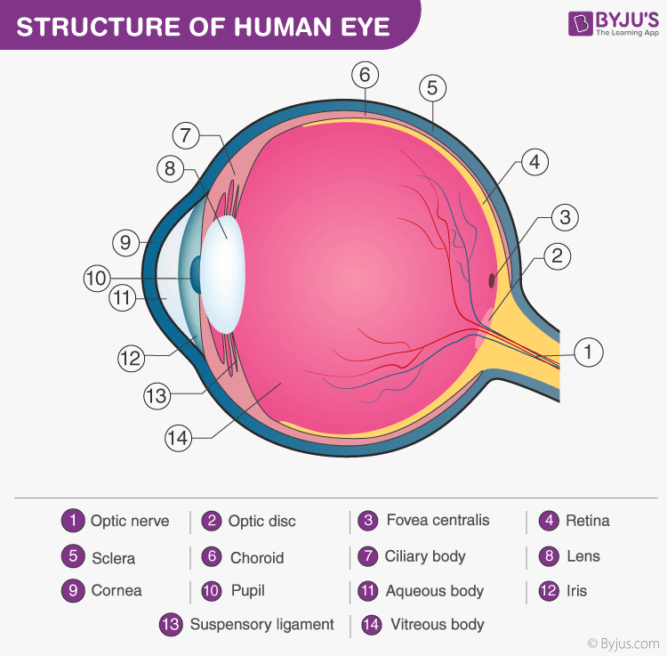
ear and eye diagram and functions look at the following descriptions and place the letter in the box where it fits eye part eye function a1pupil b1 refracts light and focuses the image on retina a2 optic nerve b2 point of central focus in the retina a3 cornea b3 hole light travels through a4 lens b4 clear outer layer protecting the eye a5 retina …
Eye and Ear Diagram.pdf -. School The City College of New York, CUNY. Course Title PHIL 10200. Uploaded By szylabombila. Pages 1. This preview shows page 1 out of 1 page. View full document. End of preview.
The ear has three parts: the outer ear, the middle ear, and inner ear. The middle ear is connected to the top of the back of the throat by the Eustachian tube which is lined with mucous, just like the inside of the nose and throat. The Eustachian tube includes the tiny bones through which sound travels to the ear.
Eye and ear diagram.
The ear is divided into three parts: Outer ear: The outer ear includes an ear canal that is is lined with hairs and glands that secrete wax.This part of the ear provides protection and channels ...
The Structure of Human Ear. Helix: It is the prominent outer rim of the external ear. Antihelix: It is the cartilage curve that is situated parallel to the helix. Crus of the Helix: It is the landmark of the outer ear, situated right above the pointy protrusion known as the tragus. Auditory Ossicles: The three small bones in the middle ear ...
Human ear. The ear is divided into three anatomical regions: the external ear, the middle ear, and the internal ear (Figure 2). The external ear is the visible portion of the ear, and it collects and directs sound waves to the eardrum. The middle ear is a chamber located within the petrous portion of the temporal bone.
of light entering the eye. Lens: The lens is a clear part of the eye behind the iris that helps to focus light, or an image, on the retina. Macula: The macula is the small, sensitive area of the retina that gives central vision. It is located in the center of the retina. Optic nerve: The optic nerve is the largest sensory nerve of the eye.
ADVERTISEMENTS: In this article we will discuss about the structure and functions of human ear. Structure of Ear: Each ear consists of three portions: (i) External ear, ADVERTISEMENTS: (ii) Middle ear and (iii) Internal ear. 1. External Ear: It comprises a pinna, external auditory meatus (canal) & tympanic membrane. (i) Pinna: ADVERTISEMENTS: The pinna is […]
Title: Eye and Ear Diagram Quiz Author: ballison Last modified by: bliss eric Created Date: 12/18/2012 1:47:00 PM Other titles: Eye and Ear Diagram Quiz
The diagram of ear is important from Class 10 and 12 perspective and is usually asked in the examinations. A brief description of the human ear along with a well-labelled diagram is given below for reference. Well-Labelled Diagram of Ear. The External ear or the outer ear consists of: Pinna/auricle is the outermost section of the ear.
The Eye and the Ear (Blank) Printable. Test students' knowledge of the human eye and ear as they color and label these diagrams.
Label Parts of the Human Ear. Select One Auditory Canal Cochlea Cochlear Nerve Eustachian Tube Incus Malleus Oval Window Pinna Round Window Semicircular Canals Stapes Tympanic Membrane Vestibular Nerve. Select One Auditory Canal Cochlea Cochlear Nerve Eustachian Tube Incus Malleus Oval Window Pinna Round Window Semicircular Canals Stapes ...
Instructions. Click the parts of the eye to see a description for each. Hover the diagram to zoom. Iris. The iris is the coloured part of the eye which surrounds the pupil. It controls light levels inside the eye, similar to the aperture on a camera. The iris contains tiny muscles that widen and narrow the pupil size.
APK Eye Diagram Review Senteo (2) PLEASE DO NOT WRITE ON THIS WORKSHEET. Directions: Select the correct letter from each word bank to identify the parts of the eye. Each term will be used only once. Word Bank #1-10: fovea centralis C. inferior rectus E. superior oblique G. superior rectus I. optic disk
Unformatted text preview: EYE DIAGRAM Please label and color the eye diagram. Optic Nerve Cornea Pupil Lens Retina Iris Write each item below from the diagram and describe its function. Cornea 1._____- It is the surface of the eye. Iris 2.
Human Eye and Ear Diagrams by Help Teaching 38 $2.00 Zip This pack features high-quality anatomical diagrams of the human eye and ear and is ideal for middle school life science or high school biology students. Included are two, one-page worksheets and answer keys.
Mnemonic Devices for the Eye and Ear By Michael A. Britt, Ph.D. Psych Test Prep and The Psych Files Hi. This is Michael Britt and I developed the mnemonic images contained in this document. I truly hope they will help you remember the various parts of the eye and ear and as a result will help you do better on your test.
anterior chamber. space between the cornea and iris/pupil. aqueous humor. clear fluid that fills the anterior chamber. lens. clear, biconvex structure posterior to the iris/pupil, focuses light entering eye. vitreous body. jelly-like substance posterior to the lens that fills the posterior 4/5 of the eye.
Take a moment to look at the ear model labeled above. This shows you all of the structures you've just learned about in the video, labeled on one diagram. Seeing them all together in this way is a great way to learn, since anatomical structures do not exist in isolation. That's why labeling the ear is an effective way to begin your revision.
THE EAR: Diagram. 1. Pinna = outer portion of ear used to collect sound waves 2. Auditory Canal = tube that sound travels down 3. Ear Drum = membrane that sends sound waves to hammer 4. Hammer = first of 3 bones. It vibrates, striking the anvil to carry on the sound 5. Anvil = gets hit by the hammer, striking the Stirrup
Study Diagrams I and II that illustrate the lens and parts of one layer of the human eye, as well as the graph below, and answer the questions that follow. 3.1 Identify parts A and B. (2) 3.2 Which Diagram (I or II) shows part of the eye… (i) where the ciliary muscles are contracted (1)
Eye Diagram Handout Author: National Eye Health Education Program of the National Eye Institute, National Institutes of Health Subject: Handout illustrating parts of the eye Keywords: parts of the eye, eye diagram, vitreous gel, iris, cornea, pupil, lens, optic nerve, macula, retina Created Date: 12/16/2011 12:39:09 PM
The fluid contained in the anterior segment of the eye. Auricle. Name the indicated structure. External auditory canal. Name the indicated structure. Outer ear. Name the indicated structure. Middle ear. Name the indicated structure.
Our eye, ear, nose and throat posters are available in paper, or lamination. Choose from normal or abnormal anatomy illustrations in a variety of sizes. Chart titles include, Glaucoma, middle ear infections, ENT, Vision and more.
Diagram of retina, approx. 400x. The final layer of the eye is the neural layer, the retina, and optic nerve. The retina has 10 layers, from outermost to innermost: 1.Pigment epithelium 2.Rod and cone processes 3.Outer limiting membrane 4.Outer nuclear layer - cell bodies of the rods and cones
The eye diagram takes its name from the fact that it has the appearance of a human eye. It is created simply by superimposing successive waveforms to form a composite image. The eye diagram is used primarily to look at digital signals for the purpose of recognizing the effects of distortion and finding its source.


0 Response to "41 eye and ear diagram"
Post a Comment