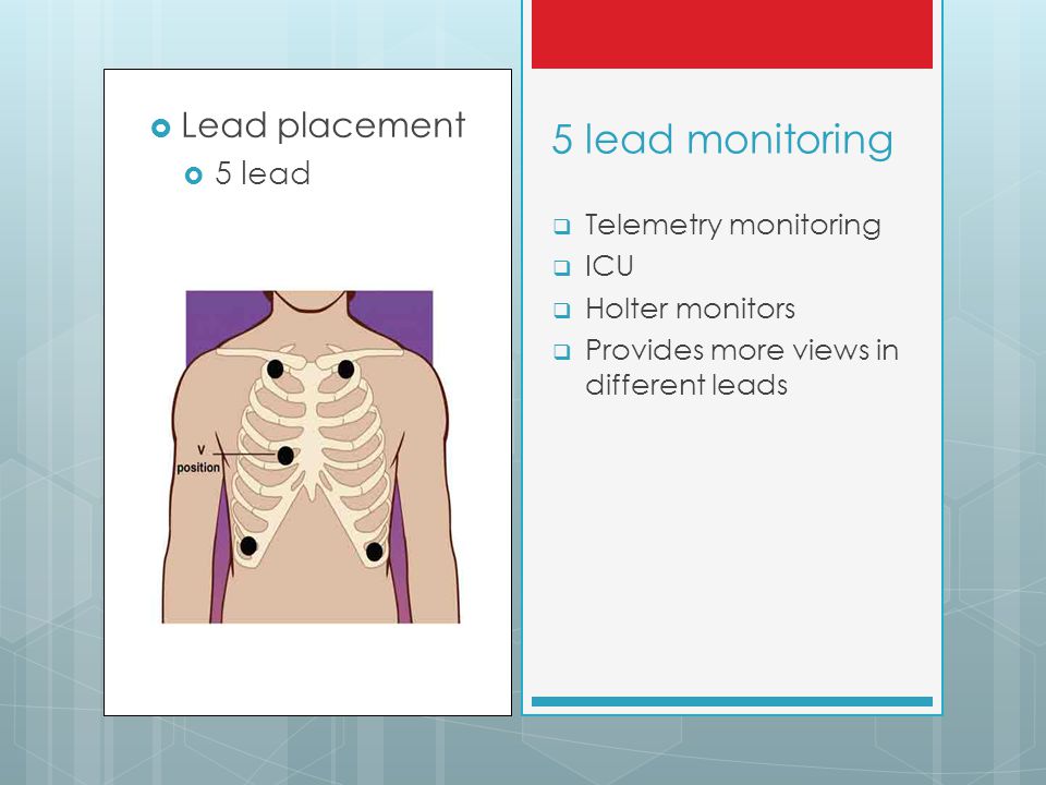
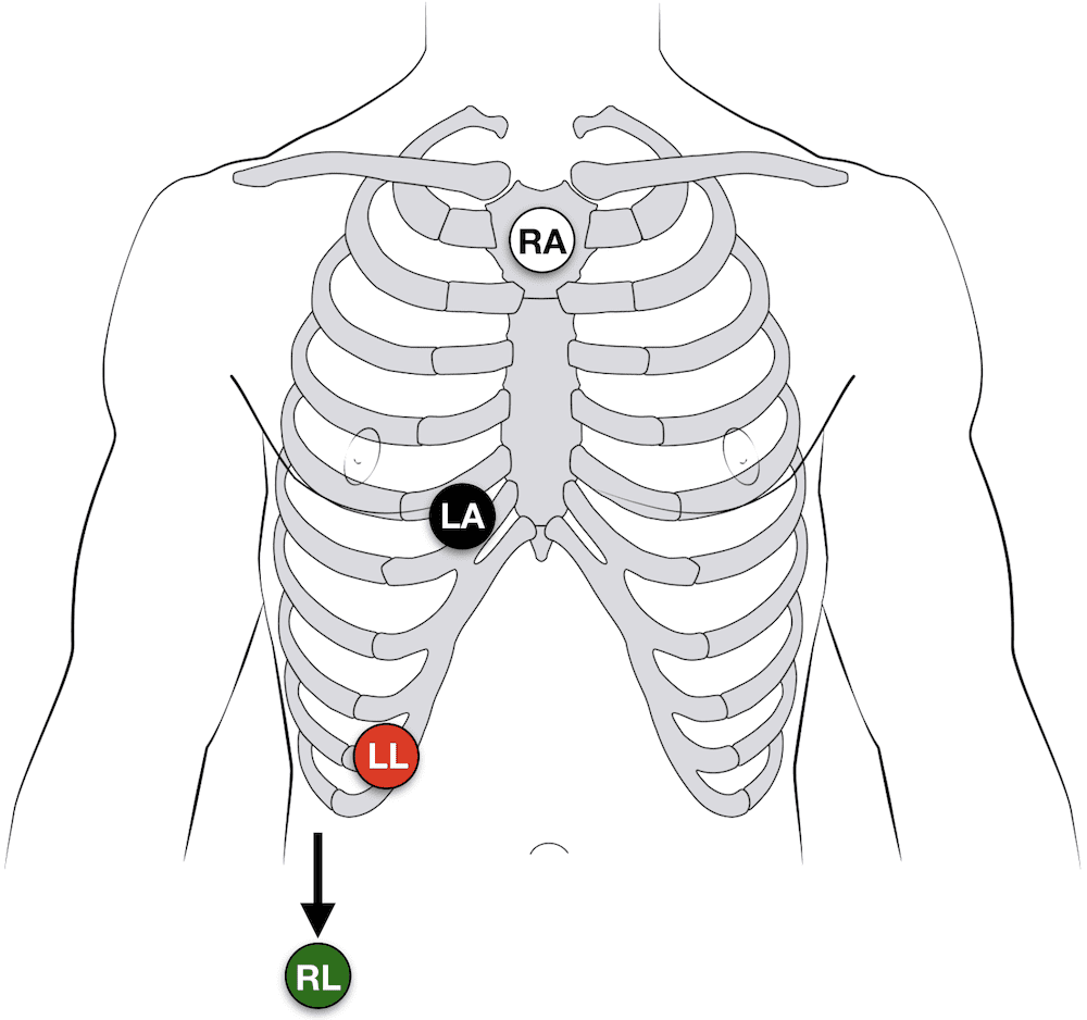
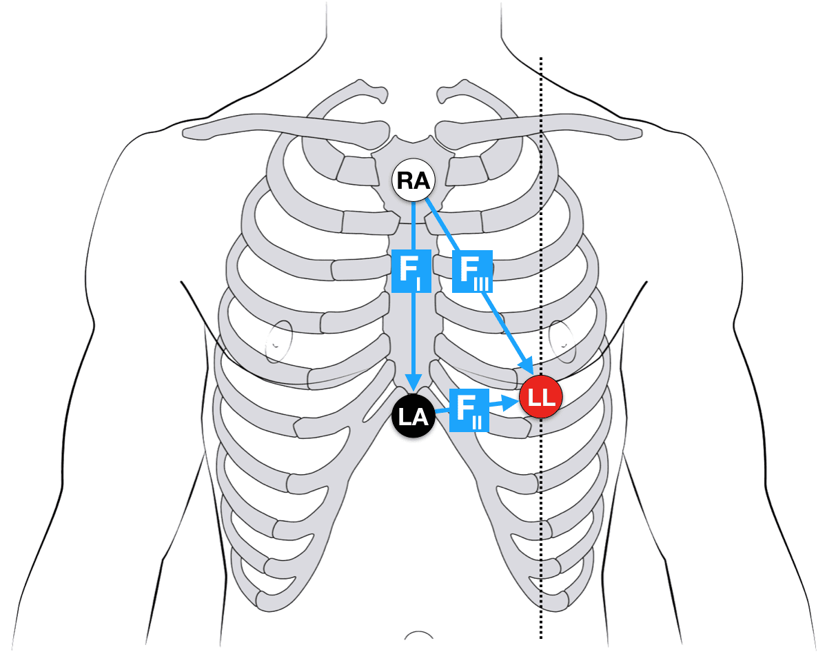
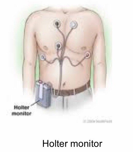
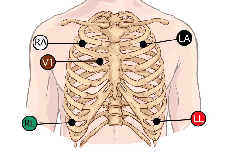
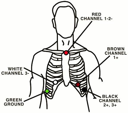
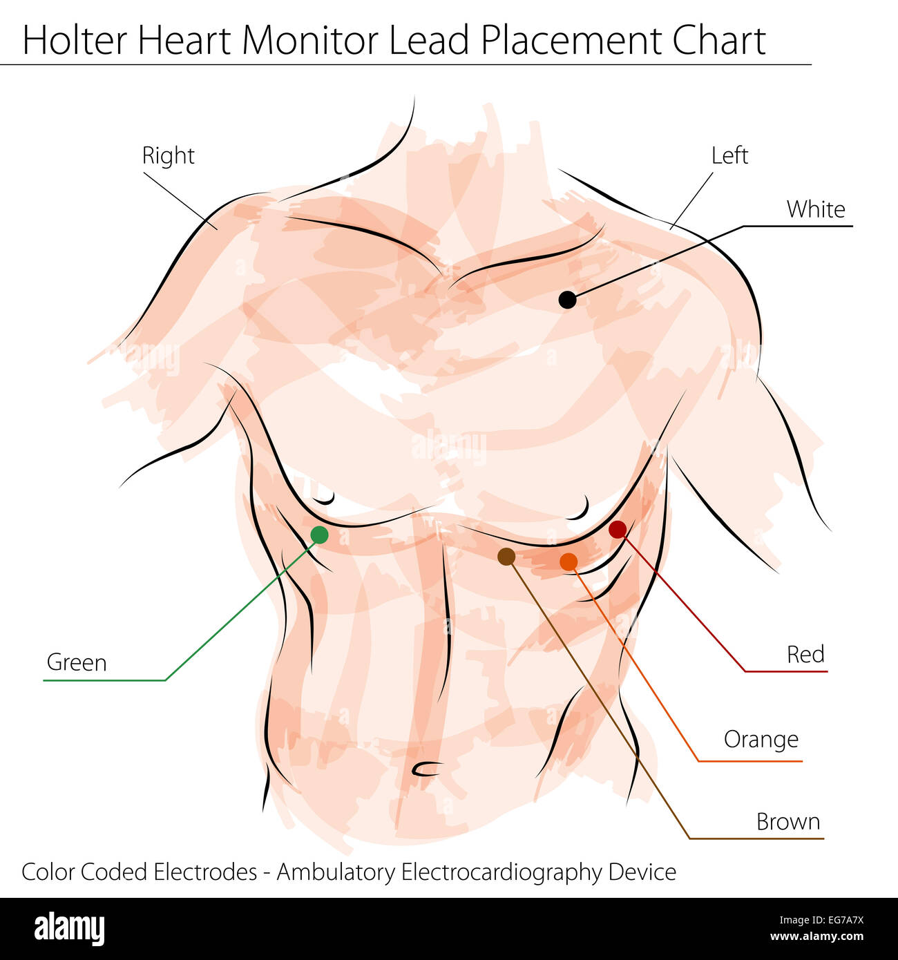
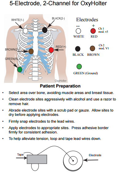
39 holter monitor 5 lead placement diagram
5-lead ECG Placement. Ok, let’s say you are on your cardiac unit and you’re admitting a patient who now needs continuous cardiac monitoring. Other names for this include placing a patient “on the monitor,” “monitored,” or on telemetry. Knowing 5 lead ECG placement is critical to a fast and efficient admission.
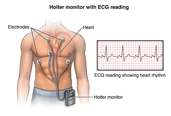
A Holter monitor is also called a portable electrocardiography (EKG) monitor. It shows your heart's electrical activity while you do your usual activities. The monitor is a small battery-operated device that you wear. It will show how fast your heart beats and if it beats in a regular pattern.
Right sided 12 lead ECG lead placement The most useful lead is V 4 R, which is obtained by placing the V4 electrode in the 5th right intercostal space in the mid-clavicular line. ST elevation in V4R has a sensitivity of 88%, specificity of 78% and diagnostic accuracy of 83% in the diagnosis of RV MI.
Holter monitor 5 lead placement diagram
• Pre-cordial lead placement – Angle of Louis –V1 – What about breast tissue? How to read an EKG • The Paper • The Waveform •The Panl. How to read an EKG • The paper – Up and down 1 box = 0.1 mV –Across 1 box = 4 ms • The rate – 10 seconds per page. How to read an EKG • The Waveform.
A Holter monitor is a battery-operated portable device that measures and records your heart’s activity continuously for 24 to 48 hours or longer depending on the type of monitoring used. The device is the size of a small camera.
•Differentiate bipolar and unipolar limb leads and precordial leads •Describe the lead placement for a 12-lead electrocardiogram •Describe the procedure for 12-lead acquisition •Describe the procedure for multi-lead acquisition using a 3-lead bipolar machine
Holter monitor 5 lead placement diagram.
Five (5) lead ECG Electrode/Cable Placement: A Five(5) lead configuration requires the placement of the three electrodes in the Three (3)-lead configuration with the addition of a fourth electrode adjacent to sternum (right side of Fourth intercostal space) and a fifth electrode on the patient's lower right abdomen.
2. Describe the significance of each ECG lead. 3. Discuss procedures for ECG electrode placement and skin preparation. 4. Discuss how the bedside monitor processes the ECG waveform for heart rate and arrhythmias, and for pacemaker detection. 5. Discuss the troubleshooting procedures for inaccurate heart rate and arrhythmia detection, and
According to ACC guidelines: 2 channel monitor (5 lead wires) White: 1st intercostal space, mid-clavicular on right Black: 1st intercostal space, mid-clavicular on left Green: Lower right at the ...
Some Holter monitors are smaller than others; the smallest are the size of a deck of cards and have four leads. They range in size up to large packs with 8+ leads. For the larger ones, you will probably either wear the battery pack/recorder around your neck, or clipped to a belt.












0 Response to "39 holter monitor 5 lead placement diagram"
Post a Comment