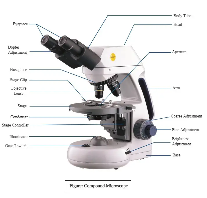
35 diagram of microscope parts
Parts of a bright-field microscope (Compound light microscope) Figure created with biorender.com It is composed of: Two lenses which include the objective lens and the eyepiece or ocular lens. Objective lens is made up of six or more glasses, which make the image clear from the object
Plant Cell Diagram 1) Cell Wall. It is the outermost, protective layer of a plant cell having a thickness of 20-80 nm. Cell walls are made up of carbohydrates such as cellulose, hemicellulose, and pectin and a complex organic polymer called lignin.
Parts of the Compound Microscope. A typical compound microscope has the following parts: 1. Head/Body: It is the upper part of the microscope. The body of the microscope holds the following parts: (a) Eyepiece: It is also called an ocular lens. It is the lens present at the top end of the metal tube.

Diagram of microscope parts
Welcome to the ultimate Microscope Quiz. This quiz will check how much do you know about Microscope Parts and Functions! The microscope has been used in science to understand elements, diseases, and cells. You must have used a microscope back in high school in the biology lab. Do you believe you understood how to use it? Take up the test and see.
Any microscope consists of three parts: Head (contains the optical parts), base ( supports the structure) and arm (connects the head to the base). The eyepiece lens is closer to the observer's eye. In comparison, the objective lens, which gives a 400 - 100 times magnified image, is closer to the object.
There are three major structural parts of a microscope: Head, Base, and Arm. Always lift a microscope by holding both the arm and base with two hands. There are ...
Diagram of microscope parts.
Illuminator: Illuminator is the most important microscope parts and it serve as light source for a microscope during slide specimen visualization. It is a continuous source of light (110 volts) used in place of a mirror. The mirror of microscope is used to reflect light from the external light source up through the bottom of the stage.
Parts of a Microscope and their Functions. The microscope comprises three main structural components: the head, the base, and the arm. HEAD: Also known as the body, the microscope's head houses the optical components in the upper portion. BASE- It serves as a support for microscopes. Illuminators for microscopic work are also included.
What are the 13 parts of a microscope? 1. Eyepiece 2. Eyepiece Tube 3. Objective Lens 4. Stage 5. Stage Clips 6. Nosepiece 7. Fine and Coarse Focus knobs 8. Illuminator 9. Aperture 10. Iris Diaphragm 11. Condenser 12. Condenser Focus Knob 13. The Rack stop Q 5. What are the 11 parts of a compound microscope?
11+ Labeled Diagram Of Electron Microscope Pics. These labeled microscope diagrams and the functions of its various parts, attempt to simplify the microscope for you. The samples are scanned in the vacuum and so they require special preparation. In addition to optical microscopes, also have your students look at electron microscopes, which ...
All of the microscopic components are held in place by the arm as well as the base. It has a light illuminator or a mirror at the base or on the nosepiece of the microscope. The nosepiece has two to five objective lenses with varying magnification power. To focus on the image, it can rotate to any position depending on the objective lens.
Print a microscope diagram, microscope worksheet, or practice microscope quiz in order to learn all the parts of a microscope.
The major goals of a microscope include magnifying the target object, produce a detailed image and making the details visible to the observer. This discussion will cover the general anatomy of light and electron microscopes, their parts, the different subtypes of each, as well as the advantages and disadvantages of each.
The structural parts of Microscope with their functions This portion of microscope is made of three important parts such as Head, base and arm. Each of these parts has a unique role. Head: The upper portion of microscope known as head. The main function of this portion is, it holds the optical elements of the unit.
Worksheet On Microscope Printable Worksheets And Activities For. Microscope Diagram For Kids Free Download On Clipartmag. Simple Microscope Definition Magnification Parts And Uses. Simple Microscope Parts Functions Diagram And Labelling. How To Draw Compound Of Microscope Easily Step By Step Youtube.
The compound microscope is an instrument that uses lenses to magnify microscopic objects and organisms. Review an introduction to the compound microscope and learn about its parts, uses, clarity ...
Parts of the optical parts are as follows: Mirror - A simple microscope has a plano-convex mirror and its primary function is to focus the surrounding light on the object being examined. Lens - The biconvex lens is placed above the stage and its function is to magnify the size of the object being examined.
Phase Contrast Microscope. This microscope was developed by Fritz Zernikes (1935), a Dutch physicist who was awarded Nobel Prize in 1953 for this contribution. It is a conventional light microscope fitted with a phase-contrast, objective, and a phase-contrast condenser.
Optical microscopes can be simple, consisting of a single lens, or compound, consisting of several optical components in line. The hand magnifying glass can ...
Parts Of a microscope. The main parts of a microscope that are easy to identify include: Head: The upper part of the microscope that houses the optical elements of the unit.; Base: The base is attached to a frame (arm) that is connected to the head of the device.The base of the microscope provides stability to the device and allows the user's hands to be free to manipulate other aspects of ...
Different Types of Microscopes and their parts and function. Microscopes are optical devices that are used to see very small objects in the laboratory. Light microscopes, electron microscopes, compound microscopes, stereo microscopes, and Digital microscopes are 5 different types of microscopes. If you are thinking of acquiring one and you are ...
Plant Cell Diagram Under Electron Microscope. It's a thin slice: Here's a diagram of a plant cell: The diagram is very clear, and labeled; but at the same time it is interpretive. Though we cannot see everything through the light microscope, some important organelles are visible and we can begin to see some structural differences.
Microscope Diagram Labeled Unlabeled And Blank Parts Of A. The 100 Lab A 3d Printable Open Source Platform For Fluorescence. Https Www Funjournal Org Wp Content Uploads 2018 09 June 17 1 S001 Pdf X89760. Compound Microscope Diagram Medical Laboratory Science Science. Microscope Quiz Worksheets Teaching Resources Tpt.
Related post: Best Microscope for Kids. Microscope Activities for Middle School (5th-8th Grade) From learning parts of a microscope and how to use one to observing and identifying the cellular components of a cross-section of a plant stem, there are a number of activities that can enrich the learning experience of middle school students.
Dec 1, 2021 — Optical parts of a microscope and their functions · Eyepiece – also known as the ocular. · Eyepiece tube – it's the eyepiece holder. · Objective ...Overview of microscope · Structural parts of a... · Optical parts of a microscope...
Microscope parts and use worksheet answer key along with labeling the parts of the microscope blank diagram available for. Can be rotated to change magnification. Parts of a eyepiece arm stageclips nosepiece focusing knobs illuminator stage objective lenses head base label the parts of the microscope.
The 16 core parts of a compound microscope are: Head (Body) Arm Base Eyepiece Eyepiece tube Objective lenses Revolving Nosepiece (Turret) Rack stop Coarse ...
admin January 4, 2021. Some of the worksheets below are Parts and Function of a Microscope Worksheets with colorful charts and diagrams to help students familiarize with the parts of the microscope along with several important questions and activities with answers. Basic Instructions. Once you find your worksheet (s), you can either click on ...
Parts of a Compound Microscope Eyepiece And Body Tube. The eyepiece is the lens through which the viewer looks to see the specimen. It usually contains a 10X or 15X power lens. The body tube connects the eyepiece to the objective lenses. Objectives and Stage Clips Objective Lenses are one of the most important parts of a Compound Microscope.
Nov 18, 2020 — Parts of a Compound Microscope · Eyepiece (ocular lens) with or without Pointer: The part that is looked through at the top of the compound ...
With Labeled Diagram and Functions How does a Compound Microscope Work? Before exploring microscope parts and functions, you should probably understand that ...
Vagina Parts: Just a Handy Diagram and Guide to the Anatomy of Your 'Down-There' Area. ... Throw a sample under a microscope and you'll also find bacteria, skin cells, and yeast spores. The ...
Let us take a look at the different parts of microscopes and their respective functions. 1. Eyepiece. it is the topmost part of the microscope. Through the eyepiece, you can visualize the object being studied. Its magnification capacity ranges between 10 and 15 times. 2.
science fair project microscope parts Biology Lessons, Science Biology, ... A diagram showing all of the parts of a compound light microscope.
Esophagus is a long hollow muscular tube extending from pharynx to stomach in animal. In esophagus histology, you will find all the layers of typical tubular organs of animal's body.. Hi dear anatomy learner, are you tired to find out the best guide to learn esophagus histology with slide images and labeled diagram? Don't worry, I have a solution for it and going to provide you a best ...
In pancreas histology of animal, you will find the both endocrine and exocrine parts. The exocrine part forms the major portion of pancreas and consists of closely packed serous acini with zymogenic cells. Again, the endocrine part consists of pancreatic islets of Langerhans which is located within the masses of serous acini.
The microscope is the most important piece of equipment in the clinic laboratory. The microscope is used to review fecal, urine, blood, and cytology samples on a daily basis. Understanding how the microscope functions, how it operates, and how to care for it will improve the reliability of your results and prolong the life of this valuable piece of equipment.

0 Response to "35 diagram of microscope parts"
Post a Comment